


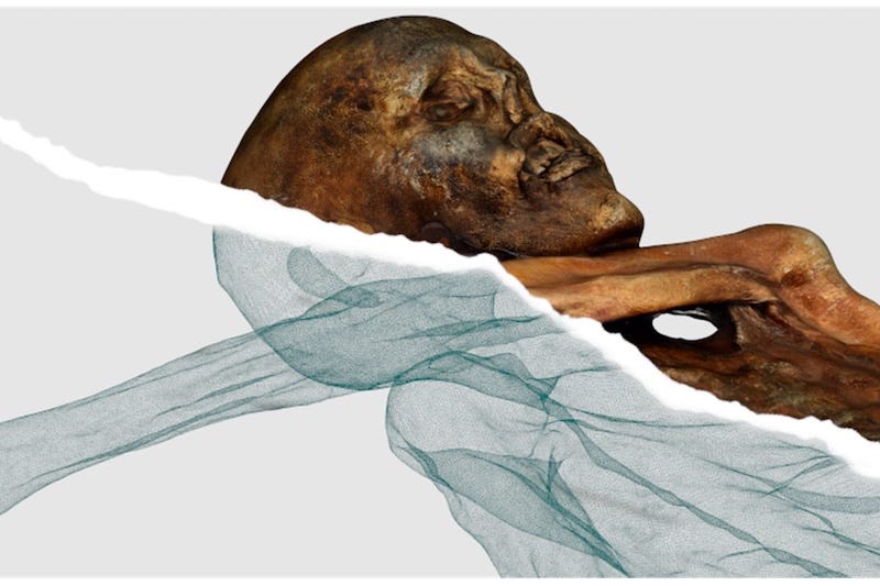
Ötzi's digital twin, with the photogrammetric skin above and the CT-derived skeleton below. Credit: South Tyrol Museum of Archaeology and Arc-Team
Ötzi is kept at minus 6.5 degrees and 99 percent humidity, in a cold cell at the South Tyrol Museum of Archaeology in Bolzano. Those numbers reproduce the glacier he came out of, and they are what stop his tissues drying out, losing mass or growing anything.
They also make him very difficult to photograph, because the conditions leave him under a protective layer of ice with wet skin beneath it, and both reflect light in a way that defeats the software used to build 3D models. Solving that problem is most of what the new paper in Heritage is about.
The work was done by Arc-Team, a small archaeological company in Cles, led by Luca Bezzi with Alessandro Bezzi, Rupert Gietl and Cicero Moraes, working with the museum, the radiology department at Bolzano hospital and a forensic pathologist from Munich. The artefacts were surveyed in 2022, the mummy in 2023 and 2024, and the paper was published on 26 August.
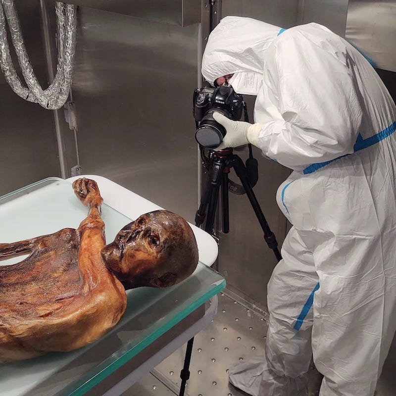
One of the three archaeologists photographing Ötzi in the sterile laboratory chamber. The shoot ran for nearly six hours. Credit: South Tyrol Museum of Archaeology and Arc-Team
Why ice is hard to photograph
Structure from Motion works by finding the same point in many overlapping photographs and calculating where it must be in space. It assumes surfaces scatter light evenly in all directions, which most things do.
Ice and wet skin do not. They reflect specularly, throwing back bright highlights that move as the camera moves. The software sees a feature that appears to be in a different place in every frame, and the reconstruction falls apart.
The team had solved this before, on a project photographing Hellenistic gold in Taranto, by working in a blacked-out room with a single polarised light source. That was not available here, because the laboratory chamber could not be darkened and its lighting could not be filtered.
So they built the light instead. A ring LED with a custom polarising filter cut from high-efficiency sheet, matched by a second polariser on the camera lens. The ring overwhelmed the ambient lighting, the crossed filters removed the glare, and what reached the sensor was diffuse reflection only.
The scan happened in a gap in the conservation schedule
Getting to him took some arranging.
The survey was slotted into a window during a microbiological inspection, when the mummy had been partially defrosted so meltwater could be sampled. That thinned the ice layer, which happened to be ideal for scanning, and the work had to finish before the routine misting with sterile water that counteracts his ongoing weight loss.
Access ran through a decontamination room into a Class 10,000 cleanroom with ultraviolet germicidal irradiation. Equipment and personnel were sterilised. Three archaeologists rotated through a shoot lasting nearly six hours, working two at a time, while the chamber was brought back down to minus 6.5 degrees.
None of them touched him. All handling was done by Oliver Peschel, a forensic pathologist from Munich, and Martina Tauber, a pathologist at Bolzano.
They took 1,294 usable photographs, 640 of the front and 654 of the back.
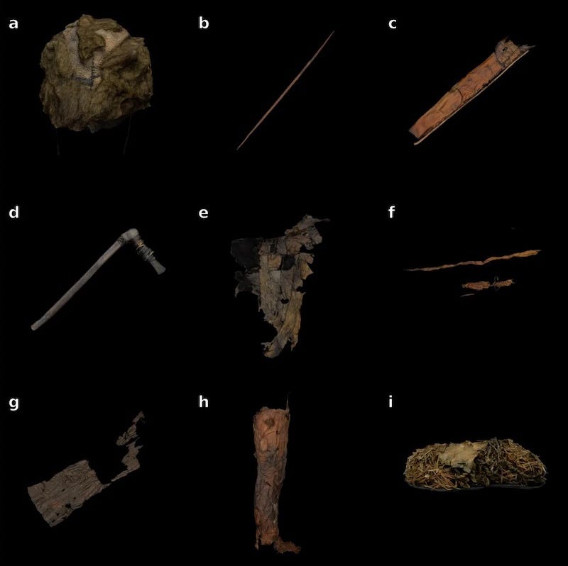
Digital models of the Iceman's clothing and equipment, from the bearskin cap at top left to a shoe at bottom right. Credit: South Tyrol Museum of Archaeology and Arc-Team
He was not in the same position for both halves
This is the part that surprised them, and it only became apparent afterwards.
Between the front survey and the back survey, his posture had changed. The right hand had moved about five centimetres and the left about one. Minimal handling, especially during the partial defrosting, is enough to shift a body that has been frozen for five thousand years.
That broke the processing pipeline, which assumes a rigid subject. The fix was to stop treating him as one object. They cut the model into four segments, the torso with head and left arm, the left forearm and hand, the upper right arm and the lower right arm, processed each separately and reassembled them.
The same problem then appeared at a larger scale. The CT scan they wanted to merge with the surface model had been taken at Bolzano hospital in 2021, and by 2023 his limbs were no longer where the scan said they were. So they took the skeleton apart digitally, disarticulating the knees, the shoulders and the joint between the skull and the first vertebra, producing five blocks that could be realigned against the new surface.
The bearskin cap needed a different technology entirely
Most of the equipment scanned adequately, the quiver, the axe, the belt and pouch, the loincloth, the shoes, the leggings, the coat and the bow.
The bearskin cap did not. Dense overlapping fur is the sort of thing photogrammetry either turns to noise or smooths into a blank surface, and neither was acceptable.
They ran the same photographs through a Neural Radiance Field instead. Rather than building a surface from points, a NeRF trains a network to answer what colour and density exist at any coordinate seen from any angle, which handles fur because it never has to decide where the surface is.
The result is two records of the same object doing different jobs. The photogrammetric model is measurable and simplified. The NeRF model is not measurable but captures what the cap actually looks like.
The reason for all of this is monitoring
The output is a web application, built on an open-source framework called 3DHOP, that lets a researcher anywhere slice the model along three axes or fade the skin away to reveal the skeleton underneath. It is the first time CT data and a photogrammetric twin have been fused this way.
But the paper is clear that the display is secondary. The plan is to repeat the survey roughly every seven years and compare the results, looking for micro-dehydration, small losses of volume from water still sublimating inside the chamber, surface damage from handling, or early signs of biological change. Quantifying that is how you find out whether the preservation regime is working before anything visible goes wrong.
There is also something CT cannot do. Tomography records density, not colour, so Ötzi’s 61 tattoos are invisible in it. They show up only in photographs.
Luca Bezzi’s framing is that the value lies in integration rather than resolution, that this is the first time the outer surface and the internal anatomy have sat in a single model. The museum’s director, Elisabeth Vallazza, puts it as documenting condition with unprecedented accuracy while keeping the object out of harm’s way.
The paper’s own phrase for it is an ethical proxy. Every future examination that happens on the model is one that does not happen on the body.
Thanks for reading. Most archaeology coverage stops at the press release. Upgrade to a paid subscription for a few dollars a month to unlock the full library of daily, evidence-based investigations, plus short documentaries and the video archive.
Sources: Bezzi, L., Bezzi, A., Gietl, R., Moraes, C., Vallazza, E., Guareschi, E.E., Tauber, M., Peschel, O., Pernter, P., and Putzer, A. (2026). “Preserving the Past, The 3D Documentation of Ötzi, the Iceman Mummy, and Its Archaeological Context.” Heritage 9(9), 339.
The five-centimeter movement of his hand is extraordinary. Intellectually, of course I understand that this is still a human body with joints and tissues rather than a fixed archaeological object, but I don't think I'd ever really considered that Ötzi can still change position.
And then having to digitally disarticulate and reposition a 5,000-year-old man's skeleton because he no longer matched his own CT scan is exactly the sort of problem I love reading about. Nobody doing that scan in 2021 was thinking, "We'd better account for Ötzi moving later."
The idea of the model as an "ethical proxy" may be my favorite part, though. Better documentation actually resulting in less handling of the original seems like exactly what conservation technology ought to accomplish.
I like the idea of "ethical proxy". It would be a fine thing if this were to be de rigeur in all future handling of uncovered human remains. Each "find'" is, after all, someone's forebear.